- Research
- Open access
- Published:
Hypothesis paper: GDF15 demonstrated promising potential in Cancer diagnosis and correlated with cardiac biomarkers
Cardio-Oncology volume 10, Article number: 56 (2024)
Abstract
Background
Cardiovascular toxicity represents a significant adverse consequence of cancer therapies, yet there remains a paucity of effective biomarkers for its timely monitoring and diagnosis. To give a first evidence able to elucidate the role of Growth Differentiation Factor 15 (GDF15) in the context of cancer diagnosis and its specific association with cardiac indicators in cancer patients, thereby testing its potential in predicting the risk of CTRCD (cancer therapy related cardiac dysfunction).
Methods
Analysis of differentially expressed genes (DEGs), including GDF15, was performed by utilizing data from the public repositories of the Cancer Genome Atlas (TCGA) and the Gene Expression Omnibus (GEO). Cardiomyopathy is the most common heart disease and its main clinical manifestations, such as heart failure and arrhythmia, are similar to those of CTRCD. Examination of GDF15 expression was conducted in various normal and cancerous tissues or sera, using available database and serum samples. The study further explored the correlation between GDF15 expression and the combined detection of cardiac troponin-T (c-TnT) and N-terminal prohormone of brain natriuretic peptide (NT-proBNP), assessing the combined diagnostic utility of these markers in predicting risk of CTRCD through longitudinal electrocardiograms (ECG).
Results
GDF15 emerged as a significant DEG in both cancer and cardiomyopathy disease models, demonstrating good diagnostic efficacy across multiple cancer types compared to healthy controls. GDF15 levels in cancer patients correlated with the established cardiac biomarkers c-TnT and NT-proBNP. Moreover, higher GDF15 levels correlated with an increased risk of ECG changes in the cancer cohort.
Conclusion
GDF15 demonstrated promising diagnostic potential in cancer identification; higher GDF15, combined with elevated cardiac markers, may play a role in the monitoring and prediction of CTRCD risk.
Introduction
Cancer and cardiovascular disease (CVD), share common risk factors such as obesity, smoking, and diabetes, and exhibit overlap in the signaling pathways that govern both normal cardiovascular physiology and tumor growth [1, 2]. The incidence of cardiovascular toxicity during or after cancer treatment has been on the rise, with heart failure (HF) being the most prevalent and severe cardiovascular complication associated with cancer therapy [3]. This trend may be attributed to improved survival rates among cancer patients, which has led to an increased prevalence of cardiomyopathy associated with aging and changes in immune function. Additionally, the cardiotoxic effects of specific cancer treatments (including chemotherapy, targeted therapy, biological agents, and irradiation) have become more pronounced [4]. Consequently, there is a pressing need to enhance the prevention, surveillance, and early management of cardiovascular diseases in patients who are at high risk of cardiac dysfunction related to cancer therapeutics throughout their treatment journey [5]. While cardiac biomarkers such as cardiac troponin-T (c-TnT) and N-terminal prohormone of brain natriuretic peptide (NT-proBNP) have been somewhat effective in guiding the initiation and monitoring of heart-protective therapy in cancer patients, there is a high demand for more sensitive and specific markers [6].
Growth Differentiation Factor 15 (GDF-15), a member of the transforming growth factor-beta (TGF-β) superfamily, is also known as macrophage inhibitory cytokine-1 (MIC-1) due to its role in inhibiting macrophage secretion of pro-inflammatory factors [7]. GDF15 is associated with a wide range of biological functions in both physiological and pathological processes, as evidenced by its alternative names [8]. Under normal conditions, GDF15 expression remains low in various tissues and serum but markedly increases in response to inflammation, tissue damage, and various disease states, including malignant tumors, CVD, diabetes, and obesity, thus acting as a stress response molecule [9,10,11,12,13]. As a diagnostic and prognostic marker for tumors, the expression of GDF15 correlates with the degree of cachexia [14]. Similarly, as a cardiovascular disease marker, it is closely related to heart failure and myocardial infarction severity [15, 16]. However, the potential of GDF15 as a predictive biomarker for CTRCD (cancer therapy related cardiac dysfunction) remains unclear, and its efficacy in assessing and monitoring cardiovascular toxicity during cancer treatment necessitates further experimental validation [17, 18].
In the current study, we identified cardiac biomarkers (including GDF15) in cancer patients using TCGA and GEO databases, and confirmed the efficiency of GDF15 in cancer identification by using serum samples, revealing the potential of GDF15 in the monitoring and predicting risk of CTRCD.
Materials and methods
Overall method framework
Recent cancer statistics indicate that approximately a quarter of all estimated cancer deaths can be attributed to digestive system tumors (DSTs) [19]. To identify cardiac biomarkers in cancer patients, we analyzed differentially expressed genes (DEGs) common to various DSTs using the TCGA database, supplemented by GEO database data related to cardiomyopathy (Figure S1). Among the five extracellular differential molecules identified across the two disease model datasets, GDF15 emerged as the most significant. Subsequent analysis revealed widespread expression of GDF15 across various normal and tumor tissues. Serum samples from 30 healthy donors and 507 cancer patients indicated that GDF15 is highly expressed in nearly all tumors, signifying significant diagnostic efficacy. Moreover, GDF15 serum levels showed a significant correlation with the cardiac markers NT-proBNP and c-TnT, particularly in cases involving acute heart failure and myocardial injury (Figure S2). An evaluation of electrocardiogram (ECG) results for cancer patients with varying GDF15 expression levels revealed a higher incidence of arrhythmic (e.g., sinus bradycardia, sinus tachycardia) and ischemic (e.g., ST changes, T-wave alterations) conditions among patients with elevated GDF15 levels, whereas patients with lower expression levels frequently exhibited normal ECG results.
Patients and healthy donors
This study enrolled a cohort comprising 30 healthy donors and 507 cancer patients treated at the Shandong Cancer Hospital and Institute from January 2022 to June 2023. The healthy donors consisted of individuals undergoing routine physical examinations, all of whom underwent serum GDF15 testing. Of these, five cases were also assessed for both c-TnT and NT-proBNP. The cancer patient group encompassed various malignancies, with 450 patients completing the full spectrum of tests for GDF15, cTnT, and NT-proBNP, and 57 liver cancer patients undergoing testing for GDF15 expression only. Comprehensive clinical data for the 450 patients across various tumor types (Table S1) were extracted from electronic medical records and the Ruimei Laboratory Information System version 6.0 (rmlis, Huangpu District, Shanghai, China), as summarized in Table 1. Informed consent was obtained from all participants prior to the study, with ethical approval granted by the Ethics Committee of the Shandong Cancer Hospital and Institute, aligning with the Declaration of Helsinki.
Data collection
Data for this research were sourced from electronic medical records and the Ruimei Laboratory Information System. The c-TnT and NT-proBNP levels in cancer patients were determined using Electrochemiluminescence on the Cobas e801 analyzer (Roche Diagnostics, GmbH, Mannheim, Germany). The normal range for NT-proBNP was considered to be 0-125 pg/ml, with < 125 pg/ml exclusionary of chronic heart failure and < 300 pg/ml exclusionary of acute heart failure. For the diagnosis of acute heart failure using NT-proBNP levels, the criteria vary by age: for individuals younger than 50 years, a level greater than 450 pg/ml is indicative; for those aged between 50 and 75 years, the threshold is above 900 pg/ml; and for those over 75 years, a level exceeding 1800 pg/ml is suggestive of acute heart failure. Additionally, for patients with a glomerular filtration rate (GFR) below 60 ml/min, a NT-proBNP level greater than 1200 pg/ml is indicative. Regarding cardiac troponin-T (c-TnT), normal levels range from 0 to 14 pg/ml. Levels between 15 and 52 pg/ml suggest myocardial injury, while levels above 52 pg/ml are indicative of acute myocardial injury. The delineation of test range and criteria, which are diagnostic criteria based on many previous studies and the long-term data accumulation of Roch company in hospital detection operation [20, 21].
Enzyme‑linked immunosorbent assay (ELISA)
Plasma samples were collected, centrifuged again at 3500 ×g for 10 min to eliminate hemocytes, including red blood cells, white blood cells, and platelets, and the supernatant was retained. The plasma levels of GDF15 from cancer patients and healthy donors were quantified using Human GDF-15 ELISA Kits (ab155432, Abcam, Cambridge, UK). Samples underwent a 10-fold dilution prior to testing, with subsequent procedures conducted as per the kit’s protocol.
Database and processing
Analysis of differentially expressed genes (DEGs) in colon adenocarcinoma (COAD), esophageal carcinoma (ESCA), liver hepatocellular carcinoma (LIHC), stomach adenocarcinoma (STAD), lung adenocarcinoma (LUAD) tissues, and adjacent tissues was conducted using the GEPIA2 online database (http://gepia2.cancer-pku.cn/#dataset). Detailed clinical data of cancer patients were obtained from The Cancer Genome Atlas (TCGA) database.
Additionally, public gene expression profiles (GSE116250) covering 14 non-failing donors (NF), 37 dilated cardiomyopathy (DCM), and 13 ischemic cardiomyopathy (ICM) were examined. DEGs identification and visualization between NF, DCM and ICM were executed through volcano plots and heatmaps. Extracellular gene analysis for protein subcellular localization utilized Hum-mPLoc 3.0.
ECG analysis
ECG data, collected on the day of or within one day before or after blood sampling, were recorded with 12-lead ECG devices and interpreted by a minimum of two cardiologists. The analysis included normal ECG readings and identification of arrhythmic (e.g., sinus bradycardia, sinus tachycardia, incomplete right bundle branch block, intraventricular block), ischemic (e.g., ST changes, T-wave alterations), and non-specific (e.g., low-voltage QRS, QT interval variations) findings.
Statistical analysis
Statistical analysis was performed using GraphPad Prism version 9.0 (GraphPad Software, CA, USA). Data were analyzed using the unpaired t-test for two groups with normal distribution, the Mann-Whitney test for non-normally distributed data, the Kruskal-Wallis test for multiple groups with non-normally distributed data, and one-way ANOVA for normally distributed datasets. Results are presented as mean ± standard deviation (SD), with a p-value of < 0.05 considered statistically significant.
Results
Screening of DEGs in TCGA database
Given the high incidence and mortality associated with digestive system tumors (DSTs), we sourced data for colon adenocarcinoma (COAD), esophageal carcinoma (ESCA), liver hepatocellular carcinoma (LIHC), and stomach adenocarcinoma (STAD) from the TCGA database, comprising a total of 1234 cancerous and 1006 non-cancerous tissue samples. The data included colon adenocarcinoma (COAD; 275 cancer vs. 349 control tissues), esophageal carcinoma (ESCA; 182 cancer vs. 286 control tissues), liver hepatocellular carcinoma (LIHC; 369 cancer vs. 160 control tissues), and stomach adenocarcinoma (STAD; 408 cancer vs. 211 control tissues), as detailed in Table S2. DEGs were determined based on a fold change > 2.0 and a P-value < 0.05, and their distribution was illustrated in volcano plots (Fig. 1A-D, Tables S3-6). An intersection of DEGs across these DSTs revealed 381 common genes (Fig. 1E, Tables S7-10), with LIHC-specific DEGs showcased in a heatmap (Fig. 1F). Given the need for biomarkers detectable in serum, we conducted a sub-localization analysis using Hum-mPLoc 3.0, identifying 48 extracellularly localized DEGs (Tables S11-14), among which GDF15 was highlighted as a significant finding (Fig. 1G).
DEGs identification in the TCGA database. Volcano plots comparing the expression fold-change of DEGs in COAD tissues (A), ESCA tissues (B), LIHC tissues (C), STAD tissues (D) compared with adjacent normal tissues. (E) Venn plot showing the shared genes among the DEGs in 4 kinds of DST tissues vs. healthy control tissues, and displayed in a heatmap (F, up-regulated marked in red or down-regulated marked in blue). (G) Shared DEGs that extracellular localized were screened and presented by heatmap (LIHC vs. normal tissues), and GDF15 was among them
Identification of DEGs clusters attributed to cardiomyopathy and DSTs patients
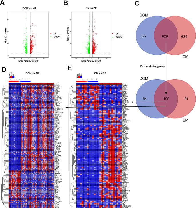
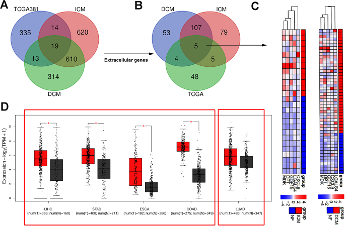
Integrating cardiomyopathy data from GSE116250, including 14 non-failing donors (NF), 37 dilated cardiomyopathy (DCM), and 13 ischemic cardiomyopathy (ICM) cases, we screened for DEGs with fold changes > 2.0 and P < 0.05, as depicted in volcano maps (Fig. 2A-B, Tables S15-16). This analysis identified 629 DEGs common between the two cardiomyopathy types (Fig. 2C). A further screen of serum biomarkers using Hum-mPLoc 3.0 revealed 105 extracellular DEGs, as displayed in heatmaps (Fig. 2D-E, Tables S17-18). A Venn diagram pinpointed 19 DEGs shared between 4 types of DST and 2 types cardiomyopathy patients, with 5 extracellular molecules identified for potential clinical blood detection, most notably GDF15 (Fig. 3B-C, Tables S19-22). The differential expression of GDF15 in lung adenocarcinoma (LUAD)/adjacent tissues was not as pronounced as in DSTs (Fig. 3D).
Screening of DEG clusters attributed to cardiomyopathy. Volcano plots comparing the expression fold-change of DEGs in dilated cardiomyopathy (DCM) vs. non-failing donors (NF) (A), and ischemic cardiomyopathy (ICM) vs. NF (B). (C) Venn plot showing the shared genes among the DEGs in DCM and ICM vs. NF, and the shared extracellular localized DEGs, presented in heatmaps (D, E); these include GDF15
Identification of DEG clusters attributed to cardiomyopathy and DST patients. Venn plot showing DEGs shared among DST patients (A), from which five differential extracellular molecules were identified (B) and are depicted in a heatmap (C). Additionally, panel (D) displays GDF15 expression levels in various tumor tissues compared to control tissues, as recorded in the TCGA database. Statistical data are expressed as mean ± standard deviation (SD), with significance determined by an unpaired two-tailed t-test: * indicates p < 0.05
GDF15 as a diagnostic biomarker for cancer patients
In pursuit of clinical insights on GDF15, we utilized integrated databases such as the Protein Atlas (https://www.proteinatlas.org/ENSG00000130513-GDF15) to examine its expression across 44 diverse tissues. This analysis, underpinned by knowledge-based annotations, utilized color-coding to demarcate tissue groups sharing functional similarities. Notably, GDF15 exhibited significant spatial expression specificity, prominently within most digestive system tissues, albeit with notable exceptions in the liver and esophagus. Immunohistochemical assays revealed distinct cytoplasmic staining patterns in colorectal and prostate cancers, among others, demonstrating the presence of GDF15. In contrast, lung cancer and certain other tumors displayed minimal to no GDF15 staining, a finding that aligns with previous analyses from the TCGA database (Fig. 4A-B). We utilized the proximity extension assay (PEA) to measure plasma concentrations of GDF15 across various cancer types. Notably, the serum levels of GDF15 in different cancer types did not always align with the patterns observed in our tissue staining (Fig. 4C). Serum samples from 30 healthy donors and 507 cancer patients representing a diverse array of cancers were analyzed to ascertain GDF15 concentrations (Table 1). In the healthy cohort (N = 30), the mean GDF15 concentration was 760.5 ± 60.30 pg/mL. In cancer patients (N = 507), however, GDF15 levels were markedly elevated, with mean concentrations ranging from 1710 ± 656.2 to 4162 ± 214.8 pg/mL (Fig. 4D). Earlier observations had indicated that normal liver tissues exhibited minimal or no GDF15 staining, whereas liver cancer tissues and serum samples displayed significantly higher GDF15 levels, the highest among all examined cancer types. Similarly, GDF15 staining was either weak or absent in both healthy and cancerous lung tissues, yet serum levels of GDF15 were considerably increased in lung cancer patients.
Expression profile of GDF15 in human normal / tumor tissues and blood. The expression of GDF15 provided by available analysis platforms in normal human tissues (A) against tumor tissues (B). Panel (C) shows the serum GDF15 levels in patients with various tumors, as reported in an online database, while panel (D) illustrates the concentrations of GDF15 that we determined in the serum of cancer patients in comparison to healthy controls. The data are expressed as mean ± standard deviation (SD), with statistical analysis conducted via an unpaired two-tailed t-test, where * signifies p < 0.05, *** signifies p < 0.001, and **** signifies p < 0.0001
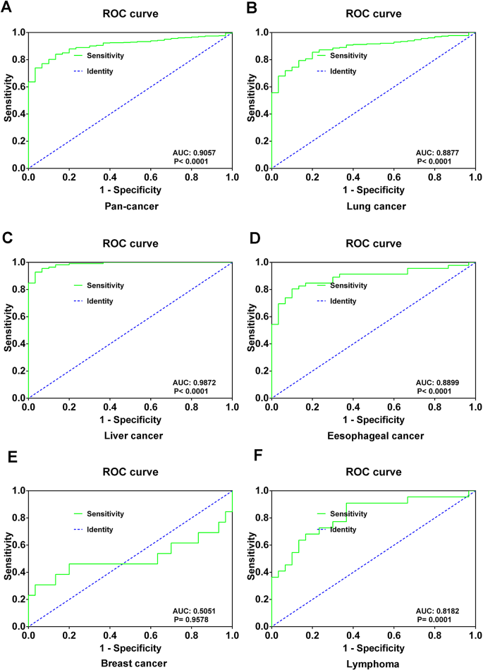
To evaluate the diagnostic utility of GDF15 for cancer, we compared serum GDF15 levels between representative cancer patient groups and healthy controls, employing receiver operating characteristic (ROC) curves. GDF15 demonstrated significant discriminative ability for pan-cancer detection, achieving an area under the curve (AUC) of 0.91 (95% confidence interval [CI] 0.8719–0.9396), with a sensitivity of 84.02% and a specificity of 86.67% compared to healthy controls (Fig. 5A). The diagnostic performance of GDF15 varied across the various cancers, exhibiting particularly strong diagnostic efficacy in liver cancer, with an AUC of 0.99 (95% confidence interval [CI] 0.9731–1.001), 92.86% sensitivity, and 96.67% specificity (Fig. 5B-F). However, in breast cancer, the diagnostic value of GDF15 was notably lower, evidenced by an AUC of 0.51 (p = 0.9578), with 30.77% sensitivity and 96.67% specificity (Fig. 5E), possibly due to the limited number of breast cancer cases (N = 13) included in the study.
Serum GDF15 levels in cancer patients correlated with cardiac indicators
Cancer therapy related cardiovascular toxicity includes myocardial injury and heart failure, immune myocarditis, hypertension, arrhythmia, coronary heart disease, venous thromboembolism, dyslipidemia. The correlation of these markers with cardiac dysfunction, as detailed in the Methods and Materials section, varies across the spectrum of potential values. Despite most c-TnT and NT-proBNP values falling within or near the normal range, with outliers presenting scattered results, it was challenging to deduce a straightforward relationship between GDF15 levels and these cardiac markers using a simple correlation analysis approach. Thus, we observed GDF15 expression across various degrees of cardiac disease in a pan-cancer context. We discovered that as c-TnT and NT-proBNP levels rose, indicating worsening cardiac disease, GDF15 expression also significantly increased (Fig. 6A-B). The mean concentration of GDF15 in individuals with NT-proBNP within the normal range (< 125 pg/ml, N = 287) was approximately 2507 ± 110.4 pg/mL. This included individuals with NT-proBNP levels below 10 pg/ml (N = 35), where GDF15 levels averaged about 1523 ± 184.4 pg/mL. When NT-proBNP levels suggested chronic heart failure (> 125 & <300 pg/ml, N = 88), GDF15 concentrations averaged around 3044 ± 226.0 pg/mL, P = 0.0230. For NT-proBNP levels indicating potential acute heart failure (> 450 pg/ml, N = 55), GDF15 levels rose to an average of 4850 ± 306.7 pg/mL, P < 0.0001. Notably, in cases with NT-proBNP exceeding 1500 pg/ml (N = 17), GDF15 levels reached a particularly high average of 5933 ± 395.4 pg/mL, P < 0.0001 (Fig. 6A).
Association Between Serum GDF15 Levels and Cardiac Biomarkers in Cancer Patients. This figure displays the correlation of serum GDF15 levels with cardiac biomarkers across all cancer types, with panels (A) and (B) illustrating GDF15 levels across varying NT-proBNP and c-TnT ranges, respectively. Panels (C) and (D) detail GDF15 levels in lung cancer patients across different NT-proBNP ranges, while panels (E) and (F) depict these levels in liver cancer patients across varying c-TnT ranges. Data represent mean ± standard error of the mean (SEM), with normal ranges for c-TnT and NT-proBNP serving as controls. N: represented the number of cases. Statistical significance was assessed using an unpaired two-tailed t-test, where * indicates p < 0.05, ** indicates p < 0.01, and **** indicates P < 0.0001
When evaluating c-TnT levels, individuals within the normal range (< 14 pg/ml, N = 366) had a mean GDF15 concentration of 2615 ± 101.1 pg/mL. This included a subset (N = 13) with c-TnT levels below 3 pg/ml, where GDF15 averaged approximately 1283 ± 277.5 pg/mL. For patients with c-TnT levels indicative of myocardial injury (15–52 pg/ml, N = 82), GDF15 levels averaged around 4303 ± 242.2 pg/mL, P < 0.0001. In cases of acute myocardial injury (c-TnT > 52 pg/ml, N = 4), GDF15 concentrations escalated to an average of 5789 ± 1151 pg/mL, P = 0.0012 (Fig. 6B).
Further analysis was conducted on lung and liver cancer patients, representing the largest subsets in this study, to more closely explore the correlation between GDF15 expression and cardiac biomarkers. Given that most c-TnT or NT-proBNP values were within or near normal ranges, with a limited number of cases showing abnormal expression, the correlation findings for these specific cancer types were not as pronounced as those observed in the broader cancer cohort. GDF15 levels were only significantly correlated with acute heart failure (NT-proBNP > 450 pg/ml) in lung cancer patients (Fig. 6C-D). In instances of myocardial injury (c-TnT > 14 pg/ml), GDF15 expression markedly increased in both lung and liver cancer (Fig. 6E-F).
The levels of serum GDF15 associated with ECG changes
Electrocardiography (ECG) has historically been used for screening and monitoring of cardiovascular toxicity caused by cancer treatments [22], however it could not be completed on all patients at the times of blood collection due to various circumstances. For instance, the patient with the highest serum level of GDF15 (7493.66 pg/ml) died the day following blood collection, leaving no ECG data. Consequently, we selected a subset of 26 patients, characterized by either relatively high or low serum GDF15 levels, for analysis and comparison of ECG results, as summarized in Table 2. The ECG findings of six representative cases are illustrated in Fig. 7. This analysis revealed that patients with lower serum levels of GDF15 generally had low levels of c-TnT or NT-proBNP, correlating with predominantly normal ECG outcomes (Fig. 7A-B). Conversely, when serum GDF15 exceeded 600 pg/ml, ECGs exhibited sinus rhythm and ST changes, despite c-TnT and NT-proBNP values remaining within normal limits (Table 2). Moreover, patients presenting with higher GDF15 expression were found to have elevated levels of c-TnT or NT-proBNP, alongside arrhythmic alterations (such as sinus bradycardia and tachycardia) and significant ischemic changes (including ST changes and T-wave alterations), suggesting a notable correlation among these biomarkers (Fig. 7C-D; Table 2). One particular case involved a patient with a high GDF15 expression (6637.38 pg/ml) whose serum levels of c-TnT (6.84 pg/ml) and NT-proBNP (16.57 pg/ml) were within normal ranges, yet the ECG revealed ST depression in leads III and avF, along with T wave inversion among other abnormal alterations (Fig. 7F; Table 2). Another significant observation was made in a patient with an exceptionally high serum GDF15 level (7493.66 pg/ml); while the c-TnT (23.03 pg/ml) and NT-proBNP (277.70 pg/ml) levels were not markedly elevated, the ECG indicated serious myocardial ischemia, as evidenced by ST segment elevation in leads II, III, avF, V5-V6 (Fig. 7E).
ECG Outcomes Related to Serum GDF15 Concentrations in Cancer Patients. This figure shows ECG findings in cancer patients, categorized by serum GDF15 levels. Panels (A) and (B) demonstrate patients with low GDF15 levels and corresponding low c-TnT and NT-proBNP levels, resulting in normal ECG outcomes. In contrast, panels (C-F) present cases with elevated GDF15 levels alongside more complex c-TnT and NT-proBNP readings, showing a heightened incidence of ECG abnormalities. All ECG recordings were conducted at a standard speed of 25 mm/s
Discussion
Cardiovascular disease and cancer are the two leading causes of death worldwide [23]. Accumulating clinical evidence demonstrates the increased risk of developing cardiac disease during cancer treatment. Cancer incidence and mortality were significantly increased in patients with heart failure [24, 25]. Addressing the cardiotoxic effects associated with anti-cancer therapies represents a formidable challenge currently confronting cardiologists and oncologists [26]. This underscores the need for a reliable serum biomarker for monitoring cardiovascular toxicity during cancer treatment. Research indicates that elevated GDF15 levels are linked to a spectrum of cardiovascular conditions, including myocardial hypertrophy, heart failure, atherosclerosis, and endothelial dysfunction. Moreover, GDF15 has been shown to precipitate cachexia and provide protection against obesity and insulin resistance in murine models [9, 27]. Notably, cardiovascular diseases, heart failure, and organ failure emerge as the prevalent clinical manifestations of cachexia induced by malignant tumors [28].
In this study, we elucidated the role of DEGs across various DSTs utilizing the TCGA database, highlighting 48 secreted proteins as potential serum biomarkers. Subsequent analyses, leveraging the GEO database, allowed us to delve into DEGs pertinent to cardiomyopathy. This dual-disease model approach culminated in the identification of five extracellular molecules, with GDF15 emerging as a notably significant biomarker for clinical detection.
Despite the variability observed in GDF15 immunohistochemical staining across normal and cancerous tissues, serum levels of GDF15 in patients with tumors significantly exceeded those in healthy controls. This disparity might be attributed to the limited sample size of healthy individuals (n = 30) and the complexity surrounding the treatment histories of cancer patients, potentially influencing GDF15 expression. Nonetheless, GDF15 demonstrated robust diagnostic utility across a spectrum of cancers, particularly standing out in DSTs where its levels were markedly elevated, aligning with findings from prior research [29].
Our results suggested an aberrant expression of GDF15 in both cancerous conditions and cardiomyopathy, posing the question of its utility as a marker for cardiac dysfunction induced by cancer treatments. While existing studies suggest GDF15’s potential as a predictive marker for CTRCD [18, 30], our analysis extends this narrative by showing a correlation between increased GDF15 levels and enhanced cardiac markers across a pan-cancer cohort. Notably, patients exhibiting the highest GDF15 serum levels also displayed elevated cardiac dysfunction markers but succumbed shortly after testing, underscoring the marker’s prognostic significance. Specifically, the patient with the highest serum GDF15 concentration (7937.295 pg/ml) presented with significantly high levels of c-TnT (342.40 pg/ml) and NT-proBNP (4348.00 pg/ml), yet died merely two days following testing. Similarly, another patient, registering extremely high GDF15 levels (7804.815 pg/ml) alongside a history of premature cardiac beats and serum levels of c-TnT (15.24 pg/ml) and NT-proBNP (1265.00 pg/ml), succumbed within a month following discharge. Through medical records review, it was found that these two patients with extremely high level of GDF15 were terminal-stage (stage IV) patients with multiple organ failure caused by bone metastasis and cachexia, which may be an important cause of their death. In contrast, when analyzing lung and liver cancer patients independently, the link between GDF15 levels and cardiac biomarkers appeared to be less pronounced. This reduced correlation suggests that the majority of these cancer patients may not exhibit pronounced cardiomyopathy post-treatment, with most cardiac indicators values falling into the normal range, except for few samples showing aberrant expressions, thereby influencing the overall statistical outcomes.
ECG has historically been critical for the diagnosis and management of cardiac injury, cardiomyopathy, and cardiovascular toxicity [22, 31, 32]. In this study, we observed the ECG patterns in patients exhibiting varying levels of GDF15 expression. Generally, we noted a correlation where elevated GDF15 levels were associated with increased c-TnT and NT-proBNP levels, alongside more pronounced arrhythmic alterations (such as sinus bradycardia) and ischemic changes (including ST segment and T-wave variations). However, there were instances where GDF15 levels were markedly high, while c-TnT and NT-proBNP levels remained within normal limits or did not show significant elevation, yet the ECG demonstrated substantial changes.
It is important to acknowledge certain limitations: not every patient had their ECG recorded in close proximity to the blood collection time, leading to disparate data without a temporally-proximal correlation. Echocardiography is common monitoring methods of CTRCD according to the Guidelines of ESC [33]. Artificial intelligence electrocardiogram served as a screening tool to detect a newly abnormal LVEF (left ventricular ejection fraction) after anthracycline-based cancer therapy [34] and lack of LVEF evaluation is indeed a limitation of this study. GDF15 has been proved to be a keen pro-inflammatory factor in many studies, and both cardiac disease and cancer have links to inflammation. The results of GDF15 and cardiac biomarkers may be the case, but whether the results can be applied accurately and specifically is certainly worth discussing. In addition, cancer subtypes and comorbidities may affect the expression of GDF15.
In conclusion, this hypothesis generating study attempts to reinforce the association between GDF15 expression and both cancer and cardiac damage after chemotherapy, underscoring its diagnostic efficacy in cancer patients and its potential in monitoring cardiovascular toxicity. While GDF15 levels generally correlate with traditional cardiac markers, the study reveals instances of discordance, suggesting a complementary role for GDF15 in the complex landscape of cancer treatment-related cardiac care.
Data availability
No datasets were generated or analysed during the current study.
References
Bertero E, Canepa M, Maack C, Ameri P. Linking heart failure to Cancer: background evidence and research perspectives. Circulation. 2018;138:735–42.
de Wit S, Glen C, de Boer RA, Lang NN. Mechanisms shared between cancer, heart failure, and targeted anti-cancer therapies. Cardiovasc Res. 2023;118:3451–66.
Otto CM. Heartbeat: heart failure induced by cancer therapy. Heart. 2019;105:1–3.
Zamorano JL, Lancellotti P, Rodriguez MD, Aboyans V, Asteggiano R, Galderisi M, et al. 2016 ESC position paper on cancer treatments and cardiovascular toxicity developed under the auspices of the ESC Committee for Practice Guidelines: the Task Force for cancer treatments and cardiovascular toxicity of the European Society of Cardiology (ESC). Eur J Heart Fail. 2017;19:9–42.
Finet JE. Management of Heart failure in Cancer patients and Cancer survivors. Heart Fail Clin. 2017;13:253–88.
Pudil R, Mueller C, Celutkiene J, Henriksen PA, Lenihan D, Dent S, et al. Role of serum biomarkers in cancer patients receiving cardiotoxic cancer therapies: a position statement from the Cardio-Oncology Study Group of the Heart Failure Association and the Cardio-Oncology Council of the European Society of Cardiology. Eur J Heart Fail. 2020;22:1966–83.
Bootcov MR, Bauskin AR, Valenzuela SM, Moore AG, Bansal M, He XY, et al. MIC-1, a novel macrophage inhibitory cytokine, is a divergent member of the TGF-beta superfamily. Proc Natl Acad Sci U S A. 1997;94:11514–9.
Assadi A, Zahabi A, Hart RA. .GDF15, an update of the physiological and pathological roles it plays: a review. Pflugers Arch. 2020;472:1535–46.
Wang D, Day EA, Townsend LK, Djordjevic D, Jorgensen SB, Steinberg GR. 2021.GDF15: emerging biology and therapeutic applications for obesity and cardiometabolic disease. Nat Rev Endocrinol 17: 592–607.
Asrih M, Wei S, Nguyen TT, Yi HS, Ryu D, Gariani K. Overview of growth differentiation factor 15 in metabolic syndrome. J Cell Mol Med. 2023;27:1157–67.
Breit SN, Brown DA, Tsai V. .GDF15 analogs as obesity therapeutics. Cell Metab. 2023;35:227–8.
Sjoberg KA, Sigvardsen CM, Alvarado-Diaz A, Andersen NR, Larance M, Seeley RJ et al. 2023.GDF15 increases insulin action in the liver and adipose tissue via a beta-adrenergic receptor-mediated mechanism. Cell Metab 35: 1327–e13405.
Wang D, Townsend LK, DesOrmeaux GJ, Frangos SM, Batchuluun B, Dumont L et al. 2023.GDF15 promotes weight loss by enhancing energy expenditure in muscle. Nature 619: 143–50.
Ling T, Zhang J, Ding F, Ma L. Role of growth differentiation factor 15 in cancer cachexia (review). Oncol Lett. 2023;26:462.
Wollert KC, Kempf T, Wallentin L. 2017.Growth differentiation factor 15 as a Biomarker in Cardiovascular Disease. Clin Chem 63: 140–51.
Sawalha K, Norgard NB, Drees BM, Lopez-Candales A. Growth differentiation factor 15 (GDF-15), a new biomarker in heart failure management. Curr Heart Fail Rep. 2023;20:287–99.
Yu LR, Cao Z, Makhoul I, Daniels JR, Klimberg S, Wei JY, et al. Immune response proteins as predictive biomarkers of doxorubicin-induced cardiotoxicity in breast cancer patients. Exp Biol Med (Maywood). 2018;243:248–55.
Ananthan K, Lyon AR. The role of biomarkers in Cardio-Oncology. J Cardiovasc Transl Res. 2020;13:431–50.
Siegel RL, Miller KD, Wagle NS, Jemal A. 2023.Cancer statistics, 2023. CA Cancer J Clin 73: 17–48.
Januzzi JJ, Chen-Tournoux AA, Moe G. Amino-terminal pro-B-type natriuretic peptide testing for the diagnosis or exclusion of heart failure in patients with acute symptoms. Am J Cardiol. 2008;101:29–38.
Hochholzer W, Morrow DA, Giugliano RP. Novel biomarkers in cardiovascular disease: update 2010. Am Heart J. 2010;160:583–94.
Zamorano JL, Lancellotti P, Rodriguez MD, Aboyans V, Asteggiano R, Galderisi M, et al. 2016 ESC position paper on cancer treatments and cardiovascular toxicity developed under the auspices of the ESC Committee for Practice Guidelines: the Task Force for cancer treatments and cardiovascular toxicity of the European Society of Cardiology (ESC). Eur Heart J. 2016;37:2768–801.
2018.Global, regional, and national age-sex-specific mortality for 282 causes of death in 195 countries and territories, 1980–2017: a systematic analysis for the global burden of Disease Study 2017. Lancet 392: 1736–88.
Camilli M, Chiabrando JG, Lombardi M, Del BM, Montone RA, Lombardo A, et al. Cancer incidence and mortality in patients diagnosed with heart failure: results from an updated systematic review and meta-analysis. Cardiooncology. 2023;9:8.
Sayour NV, Paal AM, Ameri P, Meijers WC, Minotti G, Andreadou I, et al. Heart failure pharmacotherapy and cancer: pathways and pre-clinical/clinical evidence. Eur Heart J. 2024;45:1224–40.
Fogarassy G, Vathy-Fogarassy A, Kenessey I, Veress G, Polgar C, Forster T. Preventing cancer therapy-related heart failure: the need for novel studies. J Cardiovasc Med (Hagerstown). 2021;22:459–68.
Hsu JY, Crawley S, Chen M, Ayupova DA, Lindhout DA, Higbee J, et al. Non-homeostatic body weight regulation through a brainstem-restricted receptor for GDF15. Nature. 2017;550:255–9.
Laird B, Jatoi A. Cancer cachexia: learn from yesterday, live for today and hope for tomorrow. Curr Opin Support Palliat Care. 2023;17:161.
Wang Y, Jiang T, Jiang M, Gu S. Appraising growth differentiation factor 15 as a promising biomarker in digestive system tumors: a meta-analysis. BMC Cancer. 2019;19:177.
Cartas-Espinel I, Telechea-Fernandez M, Manterola DC, Avila BA, Saavedra CN, Riffo-Campos AL. Novel molecular biomarkers of cancer therapy-induced cardiotoxicity in adult population: a scoping review. ESC Heart Fail. 2022;9:1651–65.
Valentini F, Anselmi F, Metra M, Cavigli L, Giacomin E, Focardi M, et al. Diagnostic and prognostic value of low QRS voltages in cardiomyopathies: old but gold. Eur J Prev Cardiol. 2022;29:1177–87.
Chaudhari GR, Mayfield JJ, Barrios JP, Abreau S, Avram R, Olgin JE, et al. Deep learning augmented ECG analysis to identify biomarker-defined myocardial injury. Sci Rep. 2023;13:3364.
Lyon AR, Lopez-Fernandez T, Couch LS, Asteggiano R, Aznar MC, Bergler-Klein J et al. 2022.2022 ESC guidelines on cardio-oncology developed in collaboration with the European Hematology Association (EHA), the European Society for Therapeutic Radiology and Oncology (ESTRO) and the International Cardio-Oncology Society (IC-OS). Eur Heart J 43: 4229–361.
Jacobs J, Greason G, Mangold KE, Wildiers H, Willems R, Janssens S, et al. Artificial intelligence electrocardiogram as a novel screening tool to detect a newly abnormal left ventricular ejection fraction after anthracycline-based cancer therapy. Eur J Prev Cardiol. 2024;31:560–6.
Acknowledgements
We are grateful to all the patients participated in the study and China Scholarship Council.
Funding
This work was supported by the National Natural Science Foundation of China (No. 82202836), Natural Science Foundation of Shandong Province (No. ZR202103020544), Shandong Province Medical and Health Technology Development Plan Project (No. 202002070690), Qingdao Science and Technology Demonstration and Guidance Project (22-3-7-smjk-10-nsh).
Author information
Authors and Affiliations
Contributions
Qinghai Lin designed experiments; Xiaohe Hao, Jing Kong, Xun Peng, Zhenyu Zhang and Cuiping Mao collected the laboratory results; Kun Ru, Chuanxi Zhao, Xinkai Mo and Rufei Ma provided the information of cancer patients; Qinghai Lin and Xiaohe Haoprepared Figs. 1, 2, 3, 4 and 5, Jing Kong and Rufei Ma prepared Figs. 6 and 7; Table 1, and 2. Qinghai Lin, Xiangguo Yu, Meijuan Cai and Lisheng Liu wrote the manuscript and analyzed the data; Xiaohe Hao polished the manuscript. All authors reviewed the manuscript.
Corresponding authors
Ethics declarations
Ethics approval and consent to participate
All procedures performed in studies involving human samples were reviewed and approved by the Ethics Committee of Shandong Cancer Hospital and Institute, in accordance with the Declaration of Helsinki. The participants provided their written informed consent to participate in this study.
Consent for publication
Not applicable.
Competing interests
The authors declare no competing interests.
Additional information
Publisher’s note
Springer Nature remains neutral with regard to jurisdictional claims in published maps and institutional affiliations.
Electronic supplementary material
Below is the link to the electronic supplementary material.


Rights and permissions
Open Access This article is licensed under a Creative Commons Attribution-NonCommercial-NoDerivatives 4.0 International License, which permits any non-commercial use, sharing, distribution and reproduction in any medium or format, as long as you give appropriate credit to the original author(s) and the source, provide a link to the Creative Commons licence, and indicate if you modified the licensed material. You do not have permission under this licence to share adapted material derived from this article or parts of it. The images or other third party material in this article are included in the article’s Creative Commons licence, unless indicated otherwise in a credit line to the material. If material is not included in the article’s Creative Commons licence and your intended use is not permitted by statutory regulation or exceeds the permitted use, you will need to obtain permission directly from the copyright holder. To view a copy of this licence, visit http://creativecommons.org/licenses/by-nc-nd/4.0/.
About this article
Cite this article
Hao, X., Zhang, Z., Kong, J. et al. Hypothesis paper: GDF15 demonstrated promising potential in Cancer diagnosis and correlated with cardiac biomarkers. Cardio-Oncology 10, 56 (2024). https://doi.org/10.1186/s40959-024-00263-9
Received:
Accepted:
Published:
DOI: https://doi.org/10.1186/s40959-024-00263-9


